Global Journal of Cancer Therapy
Beyond Troponins: Emerging Biomarkers for Early Detection of Chemotherapy-Induced Cardiotoxicity in Breast Cancer
2nd Department of Surgery, Aretaieio University Hospital, National and Kapodistrian University of Athens, 76 Vas. Sofias Av., 11528 Athens, Greece
Author and article information
Cite this as
Vasiliki T, et al. Beyond Troponins: Emerging Biomarkers for Early Detection of Chemotherapy-Induced Cardiotoxicity in Breast Cancer. Glob J Cancer Ther. 2026; 12(1): 4-19. Available from: 10.17352/2581-5407.000056
Copyright License
© 2026 Vasiliki T, et al. This is an open-access article distributed under the terms of the Creative Commons Attribution License, which permits unrestricted use, distribution, and reproduction in any medium, provided the original author and source are credited.Although modern breast cancer therapies have significantly improved long-term survival, reliable tools for the early detection and prediction of Cancer Therapy-Related Cardiac Dysfunction (CTRCD) remain essential. Cardiovascular toxicity encompasses a broad spectrum of manifestations, ranging from asymptomatic myocardial dysfunction to heart failure, ischemic heart disease, valvular abnormalities, arrhythmias, hypertension, and thromboembolic events.
Anthracyclines and radiation therapy are the most well-established cardiotoxic treatments. Anthracycline-induced cardiotoxicity demonstrates a dose-dependent and often irreversible pattern (Type I cardiotoxicity), primarily mediated by oxidative stress and mitochondrial injury. In contrast, HER-2 targeted therapies are associated with reversible myocardial dysfunction (Type II cardiotoxicity), mainly through the disruption of the ErbB2 signaling pathway. Newer therapies, including anti-VEGF agents and endocrine therapies combined with CDK4/6 inhibitors, further expand the spectrum of cardiovascular risks.
Current guidelines support the use of left ventricular ejection fraction, troponins, and natriuretic peptides for cardiotoxicity monitoring. However, emerging biomarkers, including myeloperoxidase, matrix metalloproteinases, galectin-3, inflammatory markers, and microRNAs, have shown potential for the early detection of myocardial injury.
This review summarizes the current evidence regarding the mechanisms, clinical features, and risk factors of cardiotoxicity associated with breast cancer therapy. It further evaluates both established and novel biomarkers and highlights the clinical value of integrating them with advanced imaging modalities to improve the early detection and monitoring of cardiac remodeling. Further research is required to validate these approaches for clinical use (Graphical abstract).
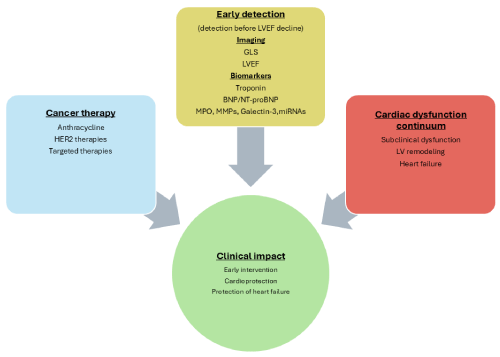
Graphical abstract: Integrated multimodal approach for the early detection of Cancer Therapy-Related Cardiac Dysfunction. Cancer therapies trigger myocardial injury through multiple mechanisms. Combined use of cardiac biomarkers and advanced imaging modalities, particularly global longitudinal strain, enables detection of subclinical dysfunction before a decline in left ventricular ejection fraction. Early identification allows for timely cardioprotective interventions and prevention of progression to heart failure.
BC: Breast Cancer; CTRCD: Cancer Therapy-Related Cardiac Dysfunction; AIC: Anthracycline-Induced Cardiotoxicity; TIC: Trastuzumab-Induced Cardiotoxicity; HF: Heart Failure; AF: Atrial Fibrillation; LVD: Left Ventricular Dysfunction; LVEF: Left Ventricular Ejection Fraction; GLS: Global Longitudinal Strain; NT-proBNP: N-terminal pro-BNP; BNP: B-type Natriuretic Peptide; cTnI and cTnT: Cardiac troponin I and Cardiac troponin T; Ais: Aromatase inhibitors; SERMs: Selective Estrogen Receptor Modulators; DOX: Doxorubicin; MACE: Major Adverse Cardiovascular Events; HER2: Human Epidermal Growth Factor Receptor 2; MPO: Myeloperoxidase; MMPs: Matrix Metalloproteinases; CRP: C-Reactive Protein; miRNAs: MicroRNAs; Gal-3: Galectin-3
Introduction
Breast cancer (BC) therapy has significantly improved the survival of a growing population of long-term cancer survivors. This progress has shifted attention toward treatment-related complications, particularly cardiovascular toxicity [1]. Cancer Therapy-Related Cardiac Dysfunction (CTRCD) encompasses a wide spectrum of clinical manifestations, ranging from asymptomatic left ventricular dysfunction (LVD) to overt heart failure (HF) [2–4].
The first anticancer treatments associated with cardiotoxicity were anthracyclines and radiation, both of which are vital tools for treating BC. Anthracycline administration causes left ventricular dysfunction (LVD) in a dose-dependent manner, whereas radiation is associated with valvular dysfunction and coronary artery disease [5]. Currently, patients undergoing targeted cancer therapies, including HER-2 inhibitors, frequently develop symptomatic HF [6].
The Cardiac Review and Evaluation Committee overseeing trastuzumab clinical trials developed one of the most precise clinical definitions of cardiotoxicity, defining drug-associated cardiotoxicity as one or more of the following: (1) a decrease in the left ventricular ejection fraction (LVEF) due to cardiomyopathy, (2) HF symptoms, (3) HF symptoms including tachycardia, S3 gallop, or both, (4) a decline in LVEF from baseline that ranges between £ 5% and < 55% with concomitant HF symptoms, or a decrease in LVEF between ³ 10% and <55% without concomitant symptoms [7]. The ESC has released Clinical Practice Guidelines to enhance the management of CTRCD. These guidelines include evaluating LVEF, determining troponin-I levels, and measuring natriuretic peptides, such as brain natriuretic peptide (BNP) [8]. Recent advances in imaging and circulating biomarkers have offered the opportunity for earlier detection of subclinical injuries. Global longitudinal strain (GLS) is the most extensively studied strain parameter, with substantial evidence supporting its diagnostic and prognostic values. GLS is the best deformation measure for identifying subclinical LVD [9]. It has become an early indicator of cardiotoxicity because conventional monitoring based on LVEF often detects myocardial dysfunction at a relatively late stage [10]. The most widely used biomarkers for identifying cardiac toxicity are cardiac troponins, which are the gold standard for identifying cardiac damage and cardiomyocyte necrosis. In asymptomatic patients, pro-B-type natriuretic peptide (proBNP) and N-terminal pro-B type natriuretic peptides (NT-proBNPs) are biomarkers of subclinical mild cardiotoxicity [11], show variable diagnostic performance and may result in inconsistent findings [12]. To lower the risk of cardiac events, predictive biomarkers for both early- and late-stage development of CTRCD are urgently needed. Clinical trials are exploring prognostic markers linked to oxidative stress, inflammation, and cardiac function to advance diagnostic, monitoring, and prognostic techniques for early- and late-stage development of CTRCD [13]. This review presents the latest findings regarding the use of novel blood biomarkers that are either already available or may be developed in the future as predictors and/or indicators of early cardiotoxicity in BC patients receiving chemotherapy (Figure 1).

Methods
We conducted this narrative review collecting data regarding BC induced cardiotoxicity based on scientific articles published on electronic databases such as PubMed and Google Scholar. The search strategy was conducted using the following keywords: (cardiotoxicity) OR (induced cardiotoxicity) OR (chemotherapy-induced cardiotoxicity) OR (cancer therapy-induced cardiotoxicity) AND (breast cancer) AND (biomarkers). The review covered studies that were published within a 26-year period, from 2000 to 2026. Our team mainly considered reviews, randomized controlled trials, cohort studies, and the reference lists of pertinent papers after eliminating articles that were not written in English. A total of 879 papers were obtained using the keyword list, 21 of them were duplicates and were eliminated. Based on the abstract’s relevancy, 379 of the 607 papers that were selected for screening were eliminated. The eligibility criteria were used to evaluate the remaining 228 papers, and 135 papers were disqualified for the reasons listed in Figure 2. This resulted in a comprehensive bibliography of 93 papers.

Results
Types of cardiotoxicity
Ewer and Lippman classified cardiotoxicity based on the type and extent of damage and the degree of reversibility. The abnormalities caused by anthracyclines in cardiac function constitute an entity, known as type 1 cardiotoxicity, which differs from the damage caused by other chemotherapeutic agents, described more recently, which differs structurally and functionally, and constitutes type II [14,15]. Risk factors that increase the likelihood of this complication have been identified and are associated with increased left ventricular end-diastolic pressure. Type I cardiotoxicity is believed to occur from the first administration of the drug, and once a threshold of damage is reached, cell death follows. The cardiovascular system can compensate for cell loss; however, severe damage remains in the patient’s myocardium, making it vulnerable to successive stresses due to infections, cardiovascular events, or other etiologies. In contrast, type II cardiotoxicity is not dose-dependent, does not appear to occur in all patients, is expressed in a wide range of severity when it occurs, and is not associated with recognizable structural abnormalities in the heart. These characteristics of type II cardiotoxicity highlight its profound contrast with those of type I cardiotoxicity. Simultaneously, the mechanism responsible for the damage is also decisively different. Type I is caused, at least in part, by oxidative stress caused by iron-based oxygen-free radicals in cardiac muscle cells. Free radicals cause peroxidation of myocyte membranes and subsequent influx of intracellular calcium. It has also been noted that there is a synergy in the function of mitochondria that seems to contribute to the appearance of these morphological changes. The mechanism of molecular damage caused by type II cardioxicity has been discovered in recent years using trastuzumab as a model. The major mechanism associated with this chemotherapeutic agent involves, at least in part, the ErbB2 pathway. Trastuzumab binds to the extracellular domain of the HER-2 protein and blocks ErbB2 signaling required for cardiomyocyte growth, repair, and survival, which helps maintain cardiac contractility, function, and structure. HER-2 activates transcription factors, such as AP-1 and nuclear factor kapa-b, which are implicated in cardiac hypertrophy and the cellular stress response. The ErbB2 molecular pathway in cardiac cells is essential for dilated cardiomyopathy induction. The main difference is that in type I, the dysfunction is considered potentially irreversible, long-term and can occur years or decades after treatment, whereas in type II, it seems to be largely reversible and short-term [15].
Anthracycline-Induced cardiotoxicity (AIC)
According to the most recent classification, which stems from the early 1980s, AIC can be classified into three distinct types: acute, which is usually reversible; occurs after a single dose or course, and the symptoms, appear within the first 14 days from the end of treatment; early onset chronic, which occurs during the first year of treatment, and is related to the appearance of a dilated-hypokinetic cardiomyopathy evolving to HF, and late-onset chronic form, developing through the years after completion of the anthracycline treatment [16].
The risk of anthracycline-induced HF is dose-dependent and increases as the cumulative dose administered increases: 3–5% with 400 mg/m2, reaching 18–48% at 700 mg/m2. Aggravating factors include age (<5 years, >65 years), previous or ongoing chest radiation, pre-existing cardiac disease, and established cardiovascular risk factors [17], as well as concomitant therapy with other agents that increase the risk, such as cyclophosphamide, paclitaxel, and trastuzumab [14].
Mechanism
The primary mechanism underlying doxorubicin (DOX)-induced cardiotoxicity is the formation of damaging reactive oxygen species under oxidative stress, which may trigger the development of cardiomyopathy. Exposure to DOX causes nicotinamide adenine dinucleotide phosphate (NAPD) oxidase to produce excessive amounts of free radicals, which can deplete antioxidant defense mechanisms over a prolonged period, resulting in cardiac myocyte apoptosis. In addition, cancer patients with impaired multidrug resistance protein (MRP) activity experience greater damage after DOX treatment, as these cell surface proteins protect cells from cytotoxic agents by transporting them out of the intracellular space [18]. All three types of cardiac cell death can be induced by DOX treatment. Apoptosis occurs through the activation of the p53 pathway, direct mitochondrial damage, and ATP depletion, which contribute to necrosis and lysosomal degradation through both iron and free radical mechanisms, resulting in autophagy [19]. Recent data indicate that the immunological response of patients may play a key role in the initiation of toxicity, which is associated with increased expression of neutrophil-specific genes and chemokines. Neutrophils, as early responders to stimuli produce cytoplasmic granules containing enzymes, such as myeloperoxidase (MPO), metalloproteinase 8 (MMP8), elastase 2 (ELA2), and cathelicidin antimicrobial peptide (CAMP) [20].
The activation of these pathways in patients with genetic variability in several HLA genes who receive doxorubicin, is not always beneficial, as they are involved in the progression of cardiovascular diseases, including atherosclerosis, thrombosis, and acute coronary syndrome [21]. Finally, neutrophil extracellular traps (NETs) represent an additional inflammatory mechanism, representing an immune response of leukocytes in which a web-like structure of DNA and intracellular contents is created. In patients with breast cancer, reactive oxygen species produced in the tumor microenvironment in the context of the inflammatory reaction stimulate the activation of this complex, a process called mitosis. NETs produce proteolytic components such as MPO and NE, provoking endothelial dysfunction, and further the release of inflammatory factors, such as IL-8 [22]. Endothelial dysfunction is associated with cardiovascular diseases, such as hypertension, coronary artery disease, chronic HF, and peripheral artery disease.
Trastuzumab-Induced Cardiotoxicity (TIC)
Human epidermal growth factor receptor 2 (HER2) is a transmembrane glycoprotein belonging to the family of receptor tyrosine kinases, which in humans includes HER1 (EGFR, ERBB1), HER2, HER3 (ERBB3), and HER4 (ERBB4). Erb receptors are involved in cell proliferation and differentiation during embryogenesis and in adulthood. Its uncontrolled overexpression is associated with the development of many types of cancer, and its overexpression is observed in 20–30% of patients with BC. In these cases, it is associated with more aggressive tumors and worse prognosis [23]. Trastuzumab was the first humanized monoclonal antibody developed and successfully used in HER-2 (+) BC. Trastuzumab binds to the extracellular domain of HER2, suppressing the intracellular signaling pathways controlled by it, preventing direct kinase inhibition to prevent molecular activation and recruitment of downstream signaling partners, and recruiting antibody-dependent cell-mediated cytotoxicity [24]. The use of targeted drugs, such as trastuzumab, pertuzumab, and more recently, tucatinib and trastuzumab teruxtecan (T-DXd), has contributed to improved survival in women with HER-2 (+) BC. Currently, early-stage BC treated with dual antibody therapy and chemotherapy achieves survival rates of more than 90% [25].
However, although survival has increased with the use of these new targeted therapies, there is no shortage of unwanted complications, the most important of which is, cardiotoxicity. This can manifest as asymptomatic, with a decrease in left ventricular ejection fraction (LVEF), or as symptomatic with the onset of congestive HF and, very rarely, cardiac death [26]. The complication rate increases in patients receiving concomitant trastuzumab and anthracycline plus cyclophosphamide and can reach up to 7% [27]. Zhang et al. (2022) demonstrated that the incidences of cardiotoxicity with trastuzumab and trastuzumab combined with pertuzumab were 14.3% and 17.9%, respectively (P > 0.05). Cardiotoxicity developed in 67 of 420 patients (15.9%), defined as a 10% decrease in LVEF of at least 10% from baseline [28].
Mechanism
Although the exact mechanism by which trastuzumab damages cardiac cells is unclear, it is thought to be associated with the critical role of erbB2 in normal cardiac function, that is, its cardioprotective role. TIC does not appear to be immune-mediated or due to the effects of the drug outside the heart. Crone et al. demonstrated in their preclinical studies that erbB2 conditional knockout mice showed increased cardiac cell apoptosis and reduced ability of the anti-apoptotic gene Bcl-xL to protect against dilated cardiomyopathy, resulting from genetic modification, while cardiomyocytes of the knockout mice had increased susceptibility to cell damage induced by anthracyclines [29]. It is worth noting that not all anti-Her-2 treatments have the same severity of damage to cardiac cells; for example, pertuzumab tends to cause milder cardiotoxicity [30]. Cardiotoxicity due to trastuzumab is partially reversible; however, treatment discontinuation is associated with disease recurrence and worse survival rates [31]. An analysis by Jacquinot et al. (2018) showed that after discontinuation or completion of trastuzumab treatment, 60.54% and 48.53% of patients failed to recover at 12 and 30 months, respectively, with an LVEF value similar to their baseline value [32]. Notably, compared with trastuzumab, pertuzumab and lapatinib have lower rates of cardiotoxicity [30,33]. Although cardiotoxicity occurs in a large proportion of patients, as mentioned above, 1/3 of them have > ΝΥΗΑ ΙI HF symptoms, less than 30% have been referred to a cardiologist, and less than 17% have received appropriate medication [34].
Other chemotherapeutic agents
In addition to the agents already mentioned, numerous other chemotherapeutic agents are used to treat BC, which have a wide range of cardiovascular side effects.
Several types of cardiovascular diseases, particularly arterial hypertension and left ventricular dysfunction (LVD), have been linked to the use of anti-VEGF agents. In addition to direct cardiomyocyte toxicity or compromised microvascular function, LVD may be associated with an elevated left ventricular afterload. They also increase the risk of myocardial ischemia and venous and arterial thromboembolic events in patients with cancer [35].
Endocrine therapy, such as aromatase inhibitors (Ais) and selective estrogen receptor modulators (SERMs) like tamoxifen (TMX), is essential for treating estrogen receptor (ER)-positive breast cancer. However, these substances cause myocardial ischemia, increased HF rates, and changes in lipid metabolism [36]. In patients with hormone receptor-positive, HER2-negative advanced BC, endocrine therapy (Ais or fulvestrant) is combined with cyclin-dependent kinase (CDK) 4/6 inhibitors, such as abemaciclib, palbociclib, and ribociclib. The addition of CDK 4/6 inhibitors to endocrine therapy increases the risk of venous thromboembolism by 3.5 times. They also cause dose-dependent QTc prolongation [37].
Over the past ten years, poly (adenine-nicotinate) (PARP) inhibitors (PARPi) have shown notable anticancer effectiveness in various tumor types that carry BRCA1/BRCA2 gene mutations, particularly in BC. However, PARPi-based treatment is associated, with a markedly elevated risk of thromboembolic events, hypertension, and MACEs [38].
Finally, the precise mechanism underlying the cardiac toxicity caused by cyclophosphamide remains to be elucidated. It has been postulated that cyclophosphamide metabolites induce oxidative stress and directly injure endothelial capillaries, leading to the extravasation of proteins, erythrocytes, and toxic metabolites. Proteins, erythrocytes, and hazardous metabolites are thought to extravasate as a result of oxidative stress and direct endothelial capillary damage caused by cyclophosphamide metabolites. Edema, interstitial bleeding, and microthrombi are immediate consequences of endothelial cell breakdown in the presence of toxic metabolites, which also damage the heart and capillary blood vessels. Clinically, these injuries present as arrhythmias and severe CF [39]. Tachyarrhythmias, hypotension, HF, myocarditis, and pericardial illness are typical symptoms that can appear up to 10 days after starting the medication, although they usually appear within the first 48 h. A rare consequence of this condition is hemorrhagic myocarditis, which is consistently and rapidly lethal [40].
Traditional biomarkers
Troponin (Tn): Cardiac troponin I (cTnI) and T (cTnT) are almost exclusively expressed in the heart and are part of the contractile machinery of myocardial cells. No reports have indicated an increase in cTnI levels after noncardiac tissue damage. The situation is more complicated for cTnT. Elevations in cTnT levels may occasionally originate from the skeletal muscle because of biochemical evidence showing that injured skeletal muscle expresses proteins that the cTnT assay may detect. According to recent evidence, these elevations may occur more frequently than previously believed in the absence of ischemic heart disease. Consequently, cTnI and cTnT are the recommended biomarkers for assessing acute and chronic myocardial damage; thus, for routine clinical use, high-sensitivity (hs)–cTn tests are advised [41]. When blood levels of cTn rise above the 99th percentile upper reference limit (URL), myocardial damage is considered to be present [Fourth Universal Definition of Myocardial Infarction, 2018]. Troponins are used in patients with cancer undergoing chemotherapy because they are a proven early sign of cardiac impairment with good diagnostic and prognostic value. Indeed, LVEF reduction and associated adverse cardiac events have been predicted by an increase in troponin levels. They are, also, strong predictors of LVEF decline and poor cardiac prognosis shortly after chemotherapy, primarily following anthracycline-containing regimens. In patients receiving trastuzumab, troponin evaluation provides a useful opportunity to identify individuals likely to develop cardiotoxicity [42]. However, TnI levels increased in approximately one-third of patients with aggressive cancers shortly after receiving high-dose chemotherapy (HDC), according to a study by Cardinale et al. (2000). The subsequent year observed a decline in left ventricular ejection fraction (LVEF), which was closely linked to the early increase. However, there is still a dearth of information on TnI behavior following this early increase and its possible influence on clinical outcomes in patients with cancer [43].
To evaluate the prognostic efficacy of cardiac troponin I for CIC in cancer patients, nine clinical studies totaling 2033 patients were included in a metanalysis of Liu et al. Compared with cTnI-negative individuals, cTnI-positive patients experienced severe cardiac events, as evidenced by a decrease in LVEF: RD = 0.279 [95% CI (0.248–0.311), p = 0.000, I2 = 81.3%, 8 trials], HF: RD = 0.117, [95% CI (0.090–0.144), p = 0.000, I2 = 77.8%, 6 trials], arrhythmias: RD = 0.057 [95% CI (0.028–0.086), p = 0.000, I2 = 0.0%, 3 trials], and cumulative events: RD = 0.318 [95% CI (0.272–0.364), p = 0.000, I2 = 73.5%, 3 trials]. Acute pulmonary edema, acute coronary syndromes, and cardiac death did not differ significantly between cTnI-positive and cTnI-negative patients [44].
Zardavas et al. (2016) found that a significant increase in the risk of LVEF drop was linked to elevated baseline troponin I (>40 ng/L) and T (>14 ng/L) levels, which were found in 56 out of 412 (13.6%) and 101 out of 407 (24.8%) patients receiving trastuzumab with chemotherapy, respectively. The researchers developed Cox regression models for the time to the occurrence of the first significant LVEF decline based on increased baseline troponin levels, because most elevations were already evident at baseline. As anticipated, these analyses yielded results comparable to the Cox analyses of troponin levels at any point in the study: for cTnI, HR = 4.52 (95% CI, 2.45 to 8.35; P<0.001) in the univariate model, and for cTnT, HR = 3.57 (95% CI, 1.95 to 6.55; P=001) in the univariate model [45].
Shafi et al. (2017) examined 82 patients with BC undergoing adjuvant or neoadjuvant chemotherapy based on anthracyclines. Eighteen (33 %) of them reported high cTnI levels. Elevated cTnI levels were observed shortly after the initial treatment round in most patients. Most patients displayed only a brief increase in cTnI, which subsided in later cycles. Therefore, after adjusting for important variables, they found that cTnI elevation was the greatest independent predictor of cardiotoxicity in multivariate analysis (95% CI, 0.003546 to 0.2535; p<.001). One cardiac event (reduction in LVEF, CHF, acute coronary syndrome, or arrhythmias) occurred in individuals with normal cTnI levels, whereas nine events occurred in patients with elevated cTnI levels [46].
Ky et al. (2014) examined eight patients every 3 months for a maximum of 15 months in a multicenter cohort of 78 patients with breast cancer receiving doxorubicin and trastuzumab therapy. Only TnI was significantly associated with the risk of cardiotoxicity when biomarker concentrations were examined 3 months after therapy initiation. The risk of future cardiotoxicity increased by nearly 40% for every 106.8 ng/l rise in the SD of TnI (HR: 1.38; 95% CI: 1.05-1.81; p= 0.020) [47].
Similar results were found in a prospective study by Demissei et al. (2020), which included 323 patients who received anthracyclines and/or trastuzumab. Their findings point to two major potential uses of hs-cTnT, particularly during the anthracycline completion phase: first, hs-cTnT levels >14 ng/L were linked to a >2-fold increased risk of CTRCD, and this threshold had a PPV of 26.5% for the prediction of CTRCD at one year after therapy completion; second, throughout the next year of follow-up, none of the patients with hs-cTnT levels <5 ng/L who had finished anthracycline therapy experienced CTRCD [48].
Natriuretic peptides
The heart ventricles release B-type natriuretic peptide (BNP) and N-terminal pro-BNP (NT-proBNP) in response to pressure and volume overloading. BNP acts on the kidneys, increasing sodium excretion, inducing diuresis, and relaxing vascular smooth muscle, thereby lowering ventricular preload. The inactive part of the cleaved pro-BNP molecule is NT-proBNP [49].
In 2020, Andersson et al. evaluated 136 patients with HER-2 positive breast cancer receiving adjuvant, neoadjuvant, or palliative chemotherapy along with HER-2 blocking. Patients underwent blood tests and echocardiography at baseline, six months, and twelve months after the initiation of chemotherapy. Five patients experienced TIC after six months, and one patient developed TIC after 12 months (cohort TIC incidence, 4.4%). NT-proBNP increased from 198.8 ± 64.0 pg/ml to 678.7 ± 132.4 pg/ml in patients with TIC (p <.05, n = 6). In contrast, at 6 months, the NT-proBNP levels of control patients decreased from 131.2 ± 20.9 pg/ml to 86.7 ± 8.8 pg/ml (p <.05, n =119) [50].
According to a retrospective observational study by Blancas et al. (2020), 27.3% of 66 patients with HER2-positive BC who received trastuzumab experienced cardiotoxicity. Cardiotoxicity was observed in 27.3% of patients receiving trastuzumab. In 66.7% of the cases, the onset of cardiac symptoms led to a diagnosis. A significant association was found between LVEF and NT-proBNP levels using the correlation test (Spearman’s coefficient = −0.156, p = 0.040). The correlation was particularly significant among women aged ≥ 50 years (−0.194, p = 0.039). A non-parametric ROC curve was utilized to assess the diagnostic validity of NT-proBNP as a biomarker for cardiotoxicity, and an ideal cut-off point of 206.5 pg/ml was determined [51].
In their study, Alves et al. included 64 BC patients who were treated with doxorubicin. CTRCD occurred in 22 patients up to one year after treatment. Three time points were monitored: before chemotherapy (T0), during the final seven days of chemotherapy (T1), and 12 months after the last doxorubicin cycle (T2). The cTnI and NT-proBNP levels in the 22 cardiotoxic patients increased by at least 20% at T1 or T2 compared to those at T0. At T1 and T2, 73% and 27% of the patients, respectively, had elevated cTnI and NT-proBNP levels. Additionally, they found that at T1 (116.55 ± 107.66 pg/mL), the NT-proBNP levels were 2.1 times higher than those at T0 (54.51 ± 28.58 pg/mL; p <.05) [52].
All 135 patients with HER2-positive BC who began trastuzumab therapy were included in a prospective, single-center cohort study by Bouwer et al. Echocardiograms were planned at regular intervals of every three months for a year to monitor heart function. NT-proBNP levels were measured at the same time points for research purposes. The trial endpoint of TIC was reached by 45 individuals (33%) at a median of 6 months. TIC was defined as LVEF <45%, an absolute decrease in LVEF >10% after enrollment, and/or the occurrence of a clinical cardiac event. Any+10 pmol/l change in NT-proBNP (at any time point during follow-up) was associated with an absolute difference in LVEF of −4.5% (95% CI -2.2% to −6.7%; p < 0.001). NT-proBNP levels and LVEF were correlated. Patients with and without TIC had mean NT-proBNP levels of 16.8 and 10.1 pmol/l, respectively. This suggests a mean difference of 6.7 pmol/l (p = 0.031). At any stage during follow-up, the HR for developing TIC was 1.06 for a +10 pmol/l change in NT-proBNP (95% CI 1.02–1.10, p = 0.008). All patients had NT-proBNP levels that were marginally higher than baseline (+2.9 pmol/l), and this difference was higher in patients with TIC (+10.2 pmol/l) than in those without TIC and was statistically significant (p = 0.037).TIC development was independently associated with NT-proBNP at baseline (HR 1.04, 95% CI 1.02–1.07; p = 0.003) and LVEF reduction during anthracycline treatment prior to starting trastuzumab (HR 1.16, 95% CI 1.07–1.25; p < 0.001). However, prior to the onset of TIC, there was no indication that NT-proBNP levels had increased gradually or abruptly. Consequently, the authors concluded that NT-proBNP is not appropriate for the early detection of TIC [53].
In their meta-analysis, Zhang et al. (2019) included four trials with 72 patients with BC who received radiation therapy. The results showed that the pooled SMD was -0.233 (95% CI, -1.113–0-.057). There was a significant difference in the pooled SMD (z=3.99, P=.000). These findings demonstrated that following radiation therapy, plasma BNP levels were clearly elevated, particularly in patients with left breast cancer, who showed no discernible change. Thus, BNP could be a biomarker of cardiac damage at high heart-absorbed doses following radiation therapy, particularly in patients with left-sided BC. [54].
Kouloubinis et al. explored the potential utility of novel biomarkers for the early detection of asymptomatic LVD in patients treated with anthracyclines. This prospective study comprised two groups of patients with BC: group A, who received chemotherapy with epirubicin for metastatic disease, and group B, who received less cardiotoxic treatment and had no residual disease. Additionally, healthy women were included for comparison purposes. In the metastatic group, plasma levels of NT-proBNP increased significantly (283.3 ± 27.2 vs. 158.0 ± 8.4 post- and pre-chemo, respectively, p < 0.001); however, the levels of NT-proBNP and other biochemical and apoptotic markers did not differ significantly between the groups, which may indicate more tissue and cellular damage [55].
In a 2019 study, Lu et al. evaluated 149 patients with BC who were treated with anthracyclines. Every 3–6 months after starting chemotherapy, the patients underwent the required cardiac checkup, which included echocardiography and a thorough physical assessment of their symptoms and indicators of heart failure. Individuals were assigned to the cardiotoxicity group if they met any of the following four criteria: LVEF <55%, ΔLVEF ≥10%, ΔLVDs ≥7 mm, or symptoms and signs of cardiac insufficiency. If not, they were assigned to the non-cardiotoxicity group. The authors discovered that the ideal diagnostic threshold of BNP following the last anthracycline chemotherapy treatment was 107.9 pg/ml, with a diagnostic sensitivity of 0.538 and a specificity of 0.794. Furthermore, the study found that the cardiotoxicity group had significantly higher serum BNP levels than the non-cardiotoxicity group prior to the second dose of chemotherapy (P=0.043), indicating a delayed decline in BNP levels as a result of established cardiac injury [56].
Novel biomarkers
Myeloperoxidase (MPO): In response to inflammatory stimuli, neutrophils travel from the bloodstream to infected tissues, where they effectively bind, ingest, and neutralize pathogens. Phagocytosed bacteria are swiftly eradicated by proteolytic enzymes, antimicrobial proteins, and reactive oxygen species. Neutrophils degranulate, releasing antimicrobial agents into the extracellular milieu and producing extracellular fibers, known as neutrophil extracellular traps (NETs), which consist of granule and nuclear components that neutralize and eliminate germs extracellularly[57]. Histones and over 30 primary and secondary granule components are proteins that bind to NETs. These include peptidoglycan-binding proteins, lactoferrin, pentraxin 3, gelatinase, elastase, myeloperoxidase, cathepsin G (CG), LL37, proteinase 3 (PR3), and other proteins with bactericidal activity that can eliminate virulence factors [58].
MPO’s primary mechanism of action involves the enzymatic conversion of hydrogen peroxide and chloride to hypochlorite, a highly effective antibacterial oxidant. HOCL initiates modification processes, such as oxidative cross-linking, nitration, and halogenation, against lipids, DNA, and lipoproteins [59]. The behavior of these circulating biomarkers may help identify individuals at a higher risk of developing cardiac injury and offer mechanistic insights into the development of AIC [60].
To ascertain whether various indicators of inflammation, hypercoagulopathy, and endothelial damage are associated with the risk of early cardiotoxicity in patients with breast cancer, Todorova et al. (2020) conducted a study of 51 patients receiving chemotherapy based on DOX. Cardiotoxicity was defined as an asymptomatic reduction in LVEF of > 10%. After controlling for confounding variables such as sex, age, BMI, and cancer type, the researchers found that the 21 patients who experienced cardiotoxicity according to the previously mentioned criteria had higher MPO levels at baseline (mean value 169.9, SD=50.7) and following the initial dosage of doxorubicin-based chemotherapy (mean value 269.6 + SD= 112.5) than patients (n=30) who did not experience a drop in LVEF. (mean value at baseline 132.6 + SD=45.6, after first treatment 174.5 + SD= 76). Their results demonstrated that elevated levels of MPO in the bloodstream prior to and during doxorubicin DOX-based therapy imply that MPO may be able to predict the risk of cardiotoxicity both before and after the initial dose of chemotherapy [61].
Blood samples obtained from 174 patients who received anthracycline-based chemotherapy in the CECCY trial were analyzed to assess changes in MPO levels during therapy. These levels were 1.3 and 1.5 times higher at 3 and 6 months (17.7 ng/mL (11.1, 31.1) and 19.2 ng/mL (11.1, 37.8)) following the start of treatment, respectively, than the baseline (13.2 ng/mL (7.9, 24.8)). However, at 3 and 6 months (16.3 ng/mL [10.3, 35.6] and 20.5 ng/mL [10.5, 37.8], respectively), following the start of anthracycline-based chemotherapy, MPO levels did not differ (p = 0.85) between patients with a decrease in LVEF by ≥10% (14.1 ng/mL [10.4, 25.5], n = 26) and those without a decrease in LVEF (18.1 ng/mL [12.3, 39.1] ), n = 148. Throughout therapy (6, 9, and 12 months), higher cTnI levels (p = 0.041), a sign of myocardial damage, were associated with higher baseline MPO levels above the median. According to the authors, higher MPO levels before chemotherapy can be used to identify patients likely to benefit from the cardioprotective effects of carvedilol (p < 0.001). In this study, cardiotoxicity was defined as a decrease in LVEF of ≥10% at any time up to six months after the end of treatment. It should be noted that the authors used similar cut-off values as previously described (350μg/L) because there were no established cut-off values for MPO, and the low incidence of cardiotoxicity observed in the CECCY trial might have made it more difficult to identify a possible link between MPO and the onset of AIC. Additionally, patients with breast cancer would presumably have a higher baseline MPO score due to increased inflammation. The MPO baseline levels in the study, however, were below the median of participants from a healthy population [60].
Lakhani et al. (2021) compared the increase in biomarker values at three and six months after the initiation of anthracycline therapy between 17 women with triple-negative BC and 17 healthy women, revealing a favorable association. At six months after starting anthracycline medication, 23% of the patients in this trial had a substantial decrease in LVEF. The decrease in ejection fraction was accompanied by an increase in TnI and MPO levels (r=0.3078) [62]. Although this study had a limited sample size, it demonstrated the potential of this biomarker in predicting cardiotoxicity and preventing its repercussions.
In another cohort study by Putt et al. (2015), eight biomarkers, including MPO, were assessed at baseline and at 3-month intervals for a maximum follow-up of 15 months in 78 patients with BC receiving doxorubicin and trastuzumab therapy. Elevated MPO levels are individually associated with a higher risk of cardiotoxicity. According to the definition provided by the Cardiac Review and Evaluation Committee, 23 women experienced > 1 cardiac event. The study found a correlation between an increase in hs-cTnI and MPO levels after three months of treatment and the potential risk of first cardiotoxicity. [HR=1.37, 95% CI (1.11-1.69), P=0.02]. At the same visit or the one that followed three months later, they also considered the correlation between each cardiovascular incident and the recurring biomarker elevations that lasted for up to 15 months after the index event. Crucially, their research indicated that an increase in MPO levels after three months continued to predict cardiotoxicity during treatment [63].
In contrast, in a study by Gullo et al., 2019, 62 patients were categorized into two groups based on cardiotoxicity, and early biomarker fold changes were examined between baseline and either 3 or 6 months. However, higher MPO levels could not differentiate individuals with cardiotoxicity (n=12) from those without (n=50) at baseline or 3 months after treatment with docetaxel, cyclophosphamide, and bevacizumab (p=0.1637). In addition, none of the biomarkers predicted later cardiotoxicity [64].
Matrix Metalloproteinases (MMPs)
Metalloproteinases (MMPs), a class of zinc-dependent endopeptidases released by tumor and stromal cells, break down the extracellular matrix (ECM) and basement membrane barriers. TIMPs, or tissue inhibitors of metalloproteinases, are particular inhibitors that control their action. According to their structure, substrate specificity, and proteolytic function, the 24 members of the MMP family that have been identified in humans are divided into subfamilies [65].
MMP-2 was proposed as a sensitive predictor of early alterations in asymptomatic patients that could develop into the cardiac clinical form, as well as a biomarker for fibrosis replacement during early remodeling. In patients with cardiac disease, MMP-9 appears to be a biomarker of severe cardiac remodeling and late-stage fibrosis [66].
Chan et al., 2021, showed for the first time that Doxy and ONO-4817, two oral MMPs inhibitors, reduce DOX-induced cardiotoxicity in vivo in mice. They found that DOX enhanced the transcription, protein levels, and activity of MMP-2 in the heart. In mouse hearts, myofilament lysis, titin proteolysis, and interstitial fibrosis were all avoided by inhibiting MMP-2 activity. DOX-impaired remodeling, systolic and diastolic function were improved by MMPs inhibitor-mediated protective effects on extracellular and intracellular matrices [67].
According to a prospective observational clinical trial by Grakova et al. (2022), patients who experienced AIC had higher serum levels of MMP2 and MMP9 by 8% (p=0.017) and 18.4% (p<0.001), respectively, than those who did not. MMP-2 levels were associated (p <0.05) with LVEF (r = − 674), end-diastolic and end-systolic dimensions (r = 0.296, r = 0.399), NT-proBNP (p = 0.568), and MMP-9 (r = 0.634) in women with AIC. In addition, LVEF (r = − 665), end-diastolic dimension (r = 0.295), end-systolic dimension (r = 0.398), and NT-proBNP (p = 0.698) all showed a significant correlation (p <0.05) with MMP-9 levels. Crucially, ROC analysis showed that both MMP-2 and MMP-9 levels might predict anthracycline-induced cardiotoxicity, although echocardiographic parameters and NT-proBNP levels did not appear to play a predictive role. Interestingly, the C/C genotype of MMP-2 and MMP-9 was associated with an adverse course of cardiotoxicity and higher levels of MMP-2 and MMP-9, whereas the T/T genotype was associated with a favorable anthracycline-induced cardiotoxicity course. Echocardiographic parameters, NT-proBNP levels, and 6-min walk test distances were analyzed. Women with the C/C genotype of MMP-2 had further progression of HF, with LVEF decreasing by 13.2%, end-systolic dimension increasing by 7.7% (p < 0.001), and end-diastolic dimension increasing by 4.0% (p < 0.001). Although there was a propensity for NT-proBNP levels to decline by 15.7% (p = 0.052) in women with the C/C genotype of MMP-9 (rs3918242), they also exhibited additional progression of heart failure, which was characterized by a drop in LVEF and an increase in LV dimensions. The authors concluded that gene polymorphisms that increase MMP-2 and MMP-9 levels in response to DOX may be used to predict cardiotoxicity risk [68].
The predictive value of MMPs at both the early and late stages of AIC in patients with BC warrants further investigation.
C-reactive protein (CRP)
C-reactive protein (CRP) is an acute-phase protein produced during inflammatory responses. In association with other cardioclinical outcomes, including myocardial infarction, congestive HF, and stable coronary artery disease, an increase in its level is predictive of diastolic dysfunction and decreased LVEF [69,70]. Hasan et al, demonstrated that using a high-sensitivity (hs) assay to measure high-sensitivity C-reactive protein (hs-CRP), BC patients were predicted to have decreased LVEF with 92.9% sensitivity and 45.7% specificity. The study’s notable alterations in hs-CRP, with a cut-off point of <5.0 mg/L (hs-CRP), imply that the risk of cardiotoxicity may vary depending on the treatment regimen and occur during treatment [71]. These findings were further supported by Onitilo et al. in a prospective study that comprised 54 patients with early-stage BC who tested positive for her-2. Because of the location of these proteins within the heart, potential indicators of cardiotoxicity, including BNP and cTnI, are likely to be unique to cardiomyopathy. CRP, on the other hand, is derived from hepatocytes and is not unique to cardiomyopathy because it is produced during a variety of inflammatory diseases, including infection. Therefore, it is not surprising that hs-CRP has a positive predictive value and a rather low specificity for reduced LVEF. When patients with BC receiving adjuvant trastuzumab therapy have elevated hs-CRP levels, which indicates the need for ongoing routine observation during trastuzumab treatment to ensure cardiac health and prevent irreversible damage, the associated cost of a false-positive (elevated hs-CRP in the absence of cardiotoxicity) would be relatively low [72]. However, the previously mentioned studies in this review, showed that patients with BC may have higher or lower CRP levels in response to treatment [61,73] and no association between CRP levels and subsequent CTRCD was observed in other trials [47].
Galectin-3 (Gal-3)
A beta-galactoside-binding lectin implicated in fibrogenesis and the inflammatory response in the failing heart is the new cardiac biomarker galectin-3 (Gal-3). It is believed that Gal-3 reflects key mechanisms underlying maladaptive cardiac remodeling. Numerous trials have shown that Gal-3 improves mortality risk prediction and ventricular remodeling in individuals with acute and chronic heart failure of different causetiologies [74]. Additionally, the median Gal-3 levels increased from baseline to therapy completion in patients undergoing anthracycline-based chemotherapy, but not in a discernible dose-dependent manner [73]. Numerous studies have been conducted to determine whether there is an association between baseline Gal-3 levels and the risk of subsequent cardiotoxicity.
For a maximum of 15 months, eight biomarkers were assessed every 3 months in a multicenter cohort of 78 BC patients who received DOX and trastuzumab therapy. Despite not being statistically significant, the observed effect sizes similar to those of TnI and MPO were linked to increases in Gal-3 levels, with an HR of 1.33 (p=0.195 for every SD increment). Furthermore, individuals undergoing chemotherapy had elevated Gal-3 levels during therapy, with a median difference between baseline and 3 months of 0.5 (-1.5 to 2.2), which was not statistically significant (p=0.14). In addition, the study found that interval changes in MPO and TnI levels could predict cardiotoxicity, with HRs of 1.36 and 1.43 per SD increase in interval change, respectively. Patients with elevated changes in both biomarkers had the highest risk of cardiotoxicity. The combination of multiple markers, such as MPO and TnI, may improve the detection of DOX and trastuzumab-induced cardiotoxicity. However, the addition of other biomarkers, such as GDF-15 or Gal-3, did not yield significant findings. Only the model containing ΔMPO retained the significance of additional biomarkers [47].
Bulten et al., 2015, used 123I-mIBG scintigraphy, conventional and strain echocardiography, and biomarker analysis one year after anthracycline treatment. On planar and SPECT 123I-mIBG scintigraphy, interobserver and intermethod variabilities were computed using washout (WO) and heart/mediastinum (H/M) ratios. Multivariate analyses were conducted to identify independent variables and correlations between 123I-mIBG scintigraphy data. The only biomarker that exhibited a significant association with the WH H/M ratio among those evaluated was Gal-3 [75].
Special attention should be paid to the systematic review and meta-analysis by Kastora et al.,2022, which included four observational studies of 1167 patients in total, with a low risk of bias. With a hazard ratio of 4.25 (95% confidence interval 3.1–5.18), doubling Gal-3 levels was associated with a higher risk of early anthracycline-induced cardiotoxicity (p < 0.0001) [76].
The results of the two final studies discussed here are inconsistent with the aforementioned conclusions. Patel et al. (2021) conducted a retrospective cohort study with 1160 patients (580 matched pairs) to compare the levels of three novel biomarkers between those who received anthracycline-based chemotherapy and those who did not. The lack of difference in Gal-3 levels between patients who received anthracycline (15.8, SD=7.5) and those who did not (16.1, SD=7.8) suggests that Gal-3 is probably not a useful biomarker of cardiotoxicity (p= 0.86) [77]. Similar findings were observed in the CECY trial, in which 174 BC patients undergoing chemotherapy with anthracyclines were randomized to assess the relationship between Gal-3 behavior and cardiotoxicity. The Gal-3 blood level trajectories of women with LVEF drop > 10% and those without LVEF drop were comparable. More precisely, baseline Gal-3 levels were observed to increase by 2- and 1.6- times at 3 months (12.4 ng/mL in the non-cardiotoxicity group and 11.0 in the cardiotoxicity group) and 6 months (10.3 ng/mL in the non-cardiotoxicity group and 10.4 in the cardiotoxicity group, respectively) relative to the baseline (6.3 ng/mL in both groups) in response to anthracycline treatment. This finding might be supported by the short-term follow-up, as the studies that demonstrated a correlation had longer follow-up [60].
MicroRNAs (miRNAs)
Non-coding RNAs with a length of 18–25 nucleotides, known as microRNAs (miRNAs), control the expression of genes by binding to mRNAs in a sequence-specific manner and causing translational inhibition or degradation of the targeted transcripts. Given that miRNAs control approximately 30% of total mRNA, they may be crucial for various physiological processes, including the control of ventricular hypertrophy and heart failure [78]. Determining whether the dynamic regulation of cardiac-expressed miRNAs may be more useful than mRNA profiling as a molecular signature for specific cardiac syndromes is of great interest because of their apparent sensitivity to clinical status and the possibility that they can be measured in peripheral blood samples (instead of myocardial biopsies). Approximately 200 of the approximately 1000 human miRNAs that have been discovered to date are expressed in the heart, and over 70% of them have been shown to be upregulated or downregulated in HF or clinical or experimental cardiac hypertrophy [79].
Seventeen women with HER-2 positive BC were assessed at baseline, three months, and six months following the initiation of chemotherapy in the study by Pillai et al. (2022). They demonstrated that cardiac troponin I exhibited a strong positive correlation with all miRNAs, including miR-34a (r = 0.3394), miR-21 (r = 0.4036), miR-133 (r = 0.5804), miR-1 (r = 0.3235), and miR-30e (r = 0.3350). Additionally, they found that cardiac troponin T was significantly positively correlated with each of the miRNAs, including miR-34a (r = 0.3882), miR-21 (r = 0.4744), miR-133 (r = 0.4242), miR-1 (r = 0.4372), and miR-30e (r = 0.4920), all at a confidence interval of >99%. In this study, compared to healthy controls, the expression of circulating miR-34a was considerably elevated six months after the initiation of trastuzumab therapy. The expression of miR-21 was significantly upregulated at 3 and 6 months after trastuzumab therapy compared to baseline and healthy controls. At six months, there was a noticeable increase in the relative expression of miR-133 compared to baseline and healthy controls. The expression of miR-1 and miR-30e was then evaluated, and after 3 and 6 months, it was significantly upregulated compared to baseline and healthy controls. The levels of any of these circulating miRNAs did not significantly change or increase between three and six months [80].
In a study by Lakhani et al. (2021), the investigators found significant upregulation of miR-34a and miR-29a in anthracycline-treated patients, which was associated with cardiac remodeling and hypertrophy. Cardiomyocyte-enriched miR-499, which has high sensitivity and specificity for myocardial injury, was also found to be a strong indicator of anthracycline-induced acute cardiac injury. Furthermore, the study found that miR-126, an early marker of heart failure, was upregulated by anthracycline treatment, possibly due to cellular damage, and miR-423 was upregulated in patients receiving anthracycline treatment, suggesting molecular changes in cardiac tissues. Additionally, their findings showed that miR-126, -423, and -499 were correlated with cardiac troponin T, whereas miR-29a, -34a, and -126 were significantly correlated with cardiac troponin I [62].
The expression level of miR-3135b in blood samples from 70 individuals with BC was assessed by Zare et al. (2022). The investigators compared 33 women with CTRCD to 37 women who received chemotherapy for BC but did not experience any adverse cardiac events. Patients with heart failure had significantly higher miR-3135b levels (P = 0.01; t-test). The standardized mean difference (SMD) was–0.57 (95% CI:–1.02 to–0.13), whereas the mean difference (MD) was–0.86 (95% CI:–1.5 to–0.19). Additionally, comparing the levels of miR-3135b in the two groups revealed that patients with heart failure had higher levels of miR-3135b than those in the control group (P < 0.05). In addition, there was a significant correlation between the ejection fraction and expression level of miR-3135b (r = 0.5, P = 0.0001). There was no significant correlation between any of the miR-3135b and either age (r = 0.03; P = 0.78), or BMI (r = 0.05; P = 0.65) [81].
In 2020, Feng et al. examined the alterations in miRNA 130a and its association with cardiotoxicity in 72 patients with HER-2 positive BC who underwent resection and were planned to receive treatment with epirubicin, cyclophosphamide, docetaxel, and trastuzumab. Cardiotoxicity was defined by any of the following circumstances:1) LVEF <35% and absolute decrease of LVEF >10, 2) heart failure, 3) acute coronary artery syndromes, and 4) fatal arrhythmia. The study found a 16.7% cumulative incidence of cardiotoxicity in patients with HER-2 BC who underwent adjuvant EC-D+T treatment. miR-130a, an miRNA targeting peroxisome proliferator-activated receptor γ, was positively correlated with cardiotoxicity. It is also linked to a higher susceptibility to hypoxic injury, cardiomyocyte apoptosis, and cardiac fibrosis. The study also found that miR-130a expression increased gradually in both cardiotoxicity and non-cardiotoxicity patients, possibly due to the production of superoxide anion free radicals and reactive oxygen species [82].
Finally, miR-4732-3p, one of the most promising cardioprotective miRNAs was found to be downregulated in blood samples of BC patients after anthracycline treatment (p < 0.05 validation study), in a prospective study by Sánchez-Sánchez et al., in 2022, who compared individuals with decreased cardiac function (n = 10) within the first year following anthracycline-based chemotherapy with individuals without cardiac dysfunction (n = 10 controls) [83].
These results further support the clinical use of our suggested panel of biomarkers and miRNAs in the evolution of CTRCD by demonstrating their ability to identify the early-onset of anthracycline-induced cardiotoxicity, even before it is detectable on echocardiography.
Other biomarkers
Inflammation and the immune system significantly influence disease and drug-induced toxicity. You et al. (2017) conducted a study in which 27 individuals were administered chemotherapy. Plasma samples were screened using multiplex immunoassays for 40 chemokines, nine matrix metalloproteinases, and 33 possible indicators of cardiovascular disease. The results demonstrated the significance of inflammation and immunity in the early subclinical response to DOX-based chemotherapy. It is likely that cardiomyocytes of immunologically sensitive patients are more vulnerable to DOX-induced injury [20].
The pathophysiology of infection, inflammation, autoimmunity, and cancer has been linked to IL-10. It is commonly recognized that IL-10 has positive effects on reducing the infiltration of inflammatory cells into the myocardium, which improves both diastolic and systolic function and reduces cardiac fibrosis. [84,85]. However, cardiac macrophages release IL-10, which has detrimental profibrotic effects on the myocardium, according to a recent in vivo model of left ventricular pressure overload. Therefore, there is disagreement over whether IL-10 causes or protects against conditions such as cancer and heart disease [86].
According to a retrospective study by Alves et al. (2022), which included 64 BC patients receiving DOX, prior to the first chemotherapy (T0), up to seven days after the last cycle ended (T1), and 12 months later (T2), the IL-10 levels of the cardiotoxicity group were significantly higher at T1 (4.96 pg/μL) than at T0 and T2 (3.77 pg/μL) (p <0.05 for both). Both the cardiotoxicity (n = 22) and non-cardiotoxicity (n = 42) groups had similar baseline cytokine levels. At T1 (p =0.006) and T2 (p =.0046), however, the cardiotoxicity group’s IL-10 levels were higher than those of the non-cardiotoxicity group. There was no difference in TNF, IL-1β, or IL-6 levels between the two groups (p >0.05). IL-10 and NT-proBNP levels at T0 and T2 were correlated with the cardiotoxicity group (r = 0.427, p =0.048; r = 0.740, p =0.004, respectively), but the non-cardiotoxicity group did not exhibit this association. Additionally, at T0, IL-10 exhibited a positive correlation with both IL-6 and TNF (r = 0.476, p =.025; r = 0.580, p =.005, respectively); however, at T1 and T2, these associations were not observed (p <0.05). Remarkably, at T1, IL-10 and IL-1β exhibited a positive correlation (r = 0.488, p =.021). IL-10 levels did not significantly correlate with either group’s cTnI, LVEF, BMI, or DOX dose (p >0.05) [52].
In a 2021 study, Micheletti et al. compared 50 healthy controls to BC patients receiving acute cycles of doxorubicin (DOX) (n = 33), paclitaxel (n = 35), or trastuzumab (n = 52) to assess inflammatory markers such as free iron, homocysteine, and TNF-α. In this study, we demonstrated that regular heart damage assessment indicators, such as CK-MB and hs-CRP, changed in the blood of patients with breast cancer following acute treatment. The same plasma samples from all treated patients concurrently showed increased levels of other pro-inflammatory mediators, such as TNF-a, homocysteine, and free iron, indicating a potential interaction between them and their association with heart injury [87] [Table 1].
Discussion
CTRCD represents a major limitation in the management of BC, particularly in the era of improved long-term survival. Early identification of subclinical myocardial injury is essential to prevent the progression to overt cardiac dysfunction and HF. This review summarizes current evidence on both established and emerging biomarkers, highlighting their role in early detection and risk stratification [88]. Traditionally, the identification of a cardiotoxicity event depends on clinical evidence, radiographic indicators (such as echocardiography or magnetic resonance imaging), or changes in biomarkers. Clinical trials have mainly reported HF and myocardial dysfunction; however, there is variability in cardiotoxic events. Recent guidelines consider a wide range of cardiac disorders of increasing significance, such as arrhythmias, valve dysfunction, coronary artery disease at any stage, arterial or venous thromboembolism, myocarditis, and pericarditis [89].
Among the available diagnostic tools, cardiac troponins combined with GLS currently represent the most robust and clinically applicable strategy for the early detection of cardiotoxicity. Troponins provide high sensitivity for myocardial injury and have demonstrated consistent prognostic value in multiple studies [44–48]. In parallel, GLS enables the detection of subclinical LVD before changes in LVEF become evident, allowing earlier clinical intervention [10].
Natriuretic peptides, including BNP and NT-proBNP, offer complementary information by reflecting myocardial wall stress and hemodynamic burden in patients with HF. However, their role in the early detection remains limited because elevations often occur at later stages of myocardial dysfunction. Despite their prognostic value, they should be interpreted in conjunction with imaging findings rather than as standalone early markers [90].
Emerging biomarkers, such as MPO, MMPs, Gal-3, inflammatory markers, and miRNAs, provide important insights into the underlying mechanisms of cardiotoxicity, including oxidative stress, inflammation, and myocardial remodeling. Although several studies suggest a potential predictive value, the current evidence remains heterogeneous and is often derived from small cohorts with variable methodologies. The absence of standardized cutoff values and limited reproducibility further restrict their integration into routine clinical practice [52,60,68,87,91].
A multimodal approach integrating circulating biomarkers with advanced imaging techniques appears to be the most effective strategy for the early detection and risk stratification of CTRCD. Several randomized multi-center prospective studies, such as CardioToX (NCT04790266) and the CardioCare project funded by Horizon 2020 (955175), are presently underway with the goal of evaluating the use of circulating biomarkers and cardiac imaging in the diagnosis of cardiotoxicity in BC patients receiving cardiotoxic cancer therapy, such as anthracyclines and/or trastuzumab [92,93]. This integrated strategy supports a shift toward personalized monitoring and timely interventions in cardio-oncology.
Limitations
The studies or trials included in this review have acknowledged several limitations. The authors’ definition of CTRCD, which clearly differs throughout the scientific literature and makes comparisons between other research difficult, is a crucial issue pertaining to any clinical investigations or trials. The diverse therapeutic regimens, the sample size of the studies/trials, the time points at which the biomarkers are evaluated, and/or the various assays employed to identify each marker may all contribute to the diversity of results shown in studies concerning several biomarkers. Additionally, it was clear that some biomarkers were not repeatable across clinical trials.
Further perspectives
The risk assessment tools while widely used to stratify cancer patients, face several validation and application problems according to recent clinical studies such as underestimation of risk and limited discriminatory power. The investigation of new biomarkers in cardio-oncology moves the emphasis from treating irreversible heart failure to preventing (CTRCD) in a proactive, precision-based manner. They enable medical professionals to intervene prior to the appearance of structural damage on routine echocardiograms. A multi-marker algorithmic approach is becoming more prevalent in the integration of these markers. Modern clinical trials are creating customized pre-treatment cardiotoxicity risk assessments by evaluating patient characteristics using machine learning algorithms. While encouraging, larger prospective studies are needed to standardize thresholds and determine the optimal monitoring intervals before these markers can reach their full potential.
Conclusion
The European Society for Medical Oncology and the European School of Cardiology have published guidelines for the prevention, diagnosis, treatment, and monitoring of individuals susceptible to cardiotoxicity. To reduce the frequency of cardiac adverse events, oncologists should adhere to these recommendations. To expand our current understanding and raise awareness regarding the biomarkers that are currently being studied for the detection and prediction of CTRCD in patients with BC, we undertook a comprehensive literature review. The advantages and disadvantages of the current leading methods for managing cardiotoxicity through biomarkers have been emphasized, with the goal of facilitating the creation of a more precise risk stratification model for predicting and early identifying cardiotoxicity in this population. We examined the existing evidence on classical biomarkers and compiled a list of recently explored biomarkers. A combination of serum and imaging biomarkers is typically advised for the best and most accurate risk assessment of those at risk of developing chemotherapy-induced cardiotoxicity. To validate the current and recently identified signs and predictors of cardiac damage, this review emphasizes the necessity of additional prospective clinical trials with adequate statistical power.
Statements
Funding
The author(s) declare that financial support was received for the research, authorship, and/or publication of this article. This work was supported by the 2nd Department of Surgery, Aretaieio University Hospital, National and Kapodistrian University of Athens, Greece.
Conflict of interest
The authors declare that the research was conducted in the absence of any commercial or financial relationships that could be construed as potential conflicts of interest.
- Totzeck M, Schuler M, Stuschke M, Heusch G, Rassaf T. Cardio-oncology: strategies for management of cancer-therapy related cardiovascular disease. Int J Cardiol. 2019 Apr 1;280:163-175. Available from: https://doi.org/10.1016/j.ijcard.2019.01.038
- Ghadri JR, Wittstein IS, Prasad A, Sharkey S, Dote K, Akashi YJ, et al. International expert consensus document on Takotsubo syndrome (Part II): diagnostic workup, outcome, and management. Eur Heart J. 2018 Jun 7;39(22):2047-2062. Available from: https://doi.org/doi:10.1093/eurheartj/ehy077
- Cartas-Espinel I, Telechea-Fernández M, Manterola Delgado C, Ávila Barrera A, Saavedra Cuevas N, Riffo-Campos AL. Novel molecular biomarkers of cancer therapy-induced cardiotoxicity in adult population: a scoping review. ESC Heart Fail. 2022 Jun;9(3):1651-1665. Available from: https://doi.org/doi:10.1002/ehf2.13735
- Lyon AR, López-Fernández T, Couch LS, Asteggiano R, Aznar MC, Bergler-Klein J, et al. 2022 ESC guidelines on cardio-oncology developed in collaboration with the European Hematology Association (EHA), the European Society for Therapeutic Radiology and Oncology (ESTRO) and the International Cardio-Oncology Society (IC-OS). Eur Heart J. 2022 Nov 1;43(41):4229-4361.Available from: https://doi.org/10.1093/eurheartj/ehac244
- Moslehi JJ. Cardiovascular toxic effects of targeted cancer therapies. N Engl J Med. 2016 Oct 13;375(15):1457-1467. Available from: https://doi.org/doi:10.1056/NEJMra1100265
- Nowsheen S, Aziz K, Park JY, Lerman A, Villarraga HR, Ruddy KJ, et al. Trastuzumab in female breast cancer patients with reduced left ventricular ejection fraction. J Am Heart Assoc. 2018 Aug 7;7(15):e008637. Available from: https://doi.org/10.1161/JAHA.118.008637
- Albini A, Pennesi G, Donatelli F, Cammarota R, De Flora S, Noonan DM. Cardiotoxicity of anticancer drugs: the need for cardio-oncology and cardio-oncological prevention. J Natl Cancer Inst. 2010 Jan 6;102(1):14-25. Available from: https://doi.org/10.1093/jnci/djp440
- Curigliano G, Cardinale D, Suter T, Plataniotis G, de Azambuja E, Sandri MT, et al. Cardiovascular toxicity induced by chemotherapy, targeted agents and radiotherapy: ESMO clinical practice guidelines. Ann Oncol. 2012 Oct;23 Suppl 7:vii155-vii166. Available from: https://doi.org/10.1093/annonc/mds293
- Liu JE, Barac A, Thavendiranathan P, Scherrer-Crosbie M. Strain imaging in cardio-oncology. JACC CardioOncol. 2020 Dec;2(5):677-689. doi:10.1016/j.jaccao.2020.10.011.
- Cardinale D, Colombo A, Bacchiani G, Tedeschi I, Meroni CA, Veglia F, et al. Early detection of anthracycline cardiotoxicity and improvement with heart failure therapy. Circulation. 2015 Jun 2;131(22):1981-1988. Available from: https://doi.org/10.1161/CIRCULATIONAHA.114.013777.0020
- Hinrichs L, Mrotzek SM, Mincu RI, Pohl J, Röll A, Michel L, et al. Troponins and natriuretic peptides in cardio-oncology patients: data from the ECoR registry. Front Pharmacol. 2020;11:740. Available from: https://doi.org/10.3389/fphar.2020.00740
- Bracun V, Aboumsallem JP, van der Meer P, de Boer RA. Cardiac biomarkers in patients with cancer: considerations, clinical implications, and future avenues. Curr Oncol Rep. 2020 Jun 9;22(7):67. Available from: https://doi.org/10.1007/s11912-020-00930-x
- Alexandraki A, Papageorgiou E, Zacharia M, Keramida K, Papakonstantinou A, Cipolla CM, et al. New insights in the era of clinical biomarkers as potential predictors of systemic therapy-induced cardiotoxicity in women with breast cancer: a systematic review. Cancers (Basel). 2023 Jun 22;15(13):3290. Available from: https://doi.org/10.3390/cancers15133290
- Bloom MW, Hamo CE, Cardinale D, Ky B, Nohria A, Baer L, et al. Cancer therapy-related cardiac dysfunction and heart failure: Part 1: definitions, pathophysiology, risk factors, and imaging. Circ Heart Fail. 2016 Jan;9(1):e002661. Available from: https://doi.org/10.1161/CIRCHEARTFAILURE.115.002661
- Ewer MS, Lippman SM. Type II chemotherapy-related cardiac dysfunction: time to recognize a new entity. J Clin Oncol. 2005 May 1;23(13):2900-2902. Available from: https://doi.org/10.1200/JCO.2005.05.827
- Cardinale D, Iacopo F, Cipolla CM. Cardiotoxicity of anthracyclines. Front Cardiovasc Med. 2020;7:26. Available from: https://doi.org/10.3389/fcvm.2020.00026
- Curigliano G, Cardinale D, Dent S, Criscitiello C, Aseyev O, Lenihan D, et al. Cardiotoxicity of anticancer treatments: epidemiology, detection, and management. CA Cancer J Clin. 2016 Jul;66(4):309-325. Available from: https://doi.org/10.3322/caac.21341
- Luu AZ, Chowdhury B, Al-Omran M, Teoh H, Hess DA, Verma S. Role of endothelium in doxorubicin-induced cardiomyopathy. JACC Basic Transl Sci. 2018 Dec;3(6):861-870. Available from: https://doi.org/10.1016/j.jacbts.2018.06.005
- Shi Y, Moon M, Dawood S, McManus B, Liu PP. Mechanisms and management of doxorubicin cardiotoxicity. Herz. 2011 Jun;36(4):296-305. Available from: https://doi.org/10.1007/s00059-011-3470-3
- Yu LR, Cao Z, Makhoul I, Daniels JR, Klimberg S, Wei JY, et al. Immune response proteins as predictive biomarkers of doxorubicin-induced cardiotoxicity in breast cancer patients. Exp Biol Med (Maywood). 2018 Feb;243(3):248-255.Available from: https://doi.org/10.1177/1535370217746383
- Gaul DS, Stein S, Matter CM. Neutrophils in cardiovascular disease. Eur Heart J. 2017 Jun 7;38(22):1702-1704. Available from: https://doi.org/10.1093/eurheartj/ehx244
- Snoderly HT, Boone BA, Bennewitz MF. Neutrophil extracellular traps in breast cancer and beyond: current perspectives on NET stimuli, thrombosis and metastasis, and clinical utility for diagnosis and treatment. Breast Cancer Res. 2019 Dec 18;21(1):145. Available from: https://doi.org/10.1186/s13058-019-1237-6
- Cho HS, Mason K, Ramyar KX, Stanley AM, Gabelli SB, Denney DW, et al. Structure of the extracellular region of HER2 alone and in complex with the Herceptin Fab. Nature. 2003 Feb 13;421(6924):756-760. Available from: https://doi.org/10.1038/nature01392
- Swain SM, Shastry M, Hamilton E. Targeting HER2-positive breast cancer: advances and future directions. Nat Rev Drug Discov. 2023 Feb;22(2):101-126. Available from: https://doi.org/10.1038/s41573-022-00579-0
- von Minckwitz G, Procter M, de Azambuja E, Zardavas D, Benyunes M, Viale G, et al. Adjuvant pertuzumab and trastuzumab in early HER2-positive breast cancer. N Engl J Med. 2017 Jul 13;377(2):122-131. Available from: https://doi.org/10.1056/NEJMoa1703643
- Eiger D, Pondé NF, Agbor-Tarh D, Moreno-Aspitia A, Piccart M, Hilbers FS, et al. Long-term cardiac outcomes of patients with HER2-positive breast cancer treated in the adjuvant lapatinib and/or trastuzumab treatment optimization trial. Br J Cancer. 2020 May;122(10):1453-1460. Available from: https://doi.org/10.1038/s41416-020-0786-x
- Seidman A, Hudis C, Pierri MK, Shak S, Paton V, Ashby M, et al. Cardiac dysfunction in the trastuzumab clinical trials experience. J Clin Oncol. 2002 Mar 1;20(5):1215-1221. Available from: https://doi.org/10.1200/JCO.2002.20.5.1215
- Zhang L, Wang Y, Meng W, Zhao W, Tong Z. Cardiac safety analysis of anti-HER2-targeted therapy in early breast cancer. Sci Rep. 2022 Aug 22;12(1):14312. Available from: https://doi.org/10.1038/s41598-022-18342-1
- Crone SA, Zhao YY, Fan L, Gu Y, Minamisawa S, Liu Y, et al. ErbB2 is essential in the prevention of dilated cardiomyopathy. Nat Med. 2002 May;8(5):459-465. Available from: https://doi.org/10.1038/nm0502-459
- Alhussein MM, Mokbel A, Cosman T, Aghel N, Yang EH, Mukherjee SD, et al. Pertuzumab cardiotoxicity in patients with HER2-positive cancer: a systematic review and meta-analysis. CJC Open. 2021 Nov;3(11):1372-1382. Available from: https://doi.org/10.1016/j.cjco.2021.06.019
- Sardesai S, Sukumar J, Kassem M, Palettas M, Stephens J, Morgan E, et al. Clinical impact of interruption in adjuvant trastuzumab therapy in patients with operable HER2-positive breast cancer. Cardiooncology. 2020 Nov 5;6(1):26. Available from: https://doi.org/10.1186/s40959-020-00081-9
- Jacquinot Q, Paget-Bailly S, Fumoleau P, Romieu G, Pierga JY, Espié M, et al. Fluctuation of the left ventricular ejection fraction in patients with HER2-positive early breast cancer treated by 12 months of adjuvant trastuzumab. Breast. 2018 Oct;41:1-7. Available from: https://doi.org/10.1016/j.breast.2018.06.001
- Perez EA, Koehler M, Byrne J, Preston AJ, Rappold E, Ewer MS. Cardiac safety of lapatinib: pooled analysis of 3689 patients enrolled in clinical trials. Mayo Clin Proc. 2008 Jun;83(6):679-686. Available from: https://doi.org/10.4065/83.6.679
- Calvillo-Argüelles O, Abdel-Qadir H, Suntheralingam S, Michalowska M, Amir E, Thavendiranathan P. Trastuzumab-related cardiotoxicity and cardiac care in patients with HER2-positive metastatic breast cancer. Am J Cardiol. 2020 Apr 15;125(8):1270-1275. Available from: https://doi.org/10.1016/j.amjcard.2020.01.029
- Pudil R, Mueller C, Čelutkienė J, Henriksen PA, Lenihan D, Dent S, et al. Role of serum biomarkers in cancer patients receiving cardiotoxic cancer therapies: a position statement from the Cardio-Oncology Study Group of the Heart Failure Association and the Cardio-Oncology Council of the European Society of Cardiology. Eur J Heart Fail. 2020 Nov;22(11):1966-1983. Available from: https://doi.org/10.1002/ejhf.2017
- Cheang I, Gue Y, Wu MZ, Huang JY, Ren QW, Chen Z, et al. Cardiovascular risks associated with adjuvant endocrine therapy in women with breast cancer: a population-based cohort study. BMC Cancer. 2025 Jul 1;25(1):1063. Available from: https://doi.org/10.1186/s12885-025-14280-z
- Lee Chuy K, Yu AF. Cardiotoxicity of contemporary breast cancer treatments. Curr Treat Options Oncol. 2019 May 9;20(6):51. Available from: https://doi.org/10.1007/s11864-019-0646-1
- Palazzo A, Ciccarese C, Iacovelli R, Cannizzaro MC, Stefani A, Salvatore L, et al. Major adverse cardiac events and cardiovascular toxicity with PARP inhibitors-based therapy for solid tumors: a systematic review and safety meta-analysis. ESMO Open. 2023 Apr;8(2):101154. Available from: https://doi.org/10.1016/j.esmoop.2023.101154
- Dhesi S, Chu MP, Blevins G, Paterson I, Larratt L, Oudit GY, et al. Cyclophosphamide-induced cardiomyopathy: a case report, review, and recommendations for management. J Investig Med High Impact Case Rep. 2013;1(1):2324709613480346. Available from: https://doi.org/10.1177/2324709613480346
- Morandi P, Ruffini PA, Benvenuto GM, La Vecchia L, Mezzena G, Raimondi R. Serum cardiac troponin I levels and ECG/Echo monitoring in breast cancer patients undergoing high-dose cyclophosphamide. Bone Marrow Transplant. 2001 Aug;28(3):277-282. Available from: https://doi.org/10.1038/sj.bmt.1703132
- Thygesen K, Alpert JS, Jaffe AS, Chaitman BR, Bax JJ, Morrow DA, et al. Fourth universal definition of myocardial infarction (2018). J Am Coll Cardiol. 2018 Oct 30;72(18):2231-2264. Available from: https://doi.org/10.1016/j.jacc.2018.08.1038
- Cardinale D, Colombo A, Torrisi R, Sandri MT, Civelli M, Salvatici M, et al. Trastuzumab-induced cardiotoxicity: clinical and prognostic implications of troponin I evaluation. J Clin Oncol. 2010 Sep 1;28(25):3910-3916. Available from: https://doi.org/10.1200/JCO.2009.27.3615
- Cardinale D, Sandri MT, Martinoni A, Tricca A, Civelli M, Lamantia G, et al. Left ventricular dysfunction predicted by early troponin I release after high-dose chemotherapy. J Am Coll Cardiol. 2000 Aug;36(2):517-522. Available from: https://doi.org/10.1016/S0735-1097(00)00748-8
- Liu Y, Liu H. Prediction of chemotherapy-mediated cardiotoxicity in patients with cancer by cardiac troponin I: a systematic review and meta-analysis. Int J Risk Saf Med. 2025 Feb;36(1):26-48. Available from: https://doi.org/10.1177/09246479241302586
- Zardavas D, Suter TM, Van Veldhuisen DJ, Steinseifer J, Noe J, Lauer S, et al. Role of troponins I and T and N-terminal prohormone of brain natriuretic peptide in monitoring cardiac safety of patients with early-stage HER2-positive breast cancer receiving trastuzumab: a Herceptin adjuvant study cardiac marker substudy. J Clin Oncol. 2017 Mar 10;35(8):878-884. Available from: https://doi.org/10.1200/JCO.2015.65.7916
- Shafi A, Siddiqui N, Imtiaz S, Din Sajid MU. Left ventricular systolic dysfunction predicted by early troponin I release after anthracycline-based chemotherapy in breast cancer patients. J Ayub Med Coll Abbottabad. 2017;29(2):266-269. Available from: https://pubmed.ncbi.nlm.nih.gov/28718245/
- Ky B, Putt M, Sawaya H, French B, Januzzi JL, Sebag IA, et al. Early increases in multiple biomarkers predict subsequent cardiotoxicity in patients with breast cancer treated with doxorubicin, taxanes, and trastuzumab. J Am Coll Cardiol. 2014 Mar 4;63(8):809-816. Available from: https://doi.org/10.1016/j.jacc.2013.10.061
- Demissei BG, Hubbard RA, Zhang L, Smith AM, Sheline K, McDonald C, et al. Changes in cardiovascular biomarkers with breast cancer therapy and associations with cardiac dysfunction. J Am Heart Assoc. 2020 Jan 21;9(2):e014708. Available from: https://doi.org/10.1161/JAHA.119.014708
- Taylor CJ, Roalfe AK, Iles R, Hobbs FDR. The potential role of NT-proBNP in screening for and predicting prognosis in heart failure: a survival analysis. BMJ Open. 2014 Apr 1;4(4):e004675. Available from: https://doi.org/10.1136/BMJOPEN-2013-004675
- Andersson AE, Linderholm B, Giglio D. Delta NT-proBNP predicts cardiotoxicity in HER2-positive breast cancer patients treated with trastuzumab. Acta Oncol. 2021 Apr;60(4):475-481. Available from: https://doi.org/10.1080/0284186X.2020.1842904
- Blancas I, Martín-Pérez FJ, Garrido JM, Rodríguez-Serrano F. NT-proBNP as predictor factor of cardiotoxicity during trastuzumab treatment in breast cancer patients. Breast. 2020 Dec;54:106-113. Available from: https://doi.org/10.1016/j.breast.2020.09.001
- Alves MT, Simões R, Pestana RMC, de Oliveira AN, Oliveira HHM, Soares CE, et al. Interleukin-10 levels are associated with doxorubicin-related cardiotoxicity in breast cancer patients in a one-year follow-up study. Immunol Invest. 2022 May;51(4):883-898. Available from: https://doi.org/10.1080/08820139.2021.1882486
- Bouwer NI, Liesting C, Kofflard MJM, Sprangers-van Campen SM, Brugts JJ, Kitzen JJEM, et al. NT-proBNP correlates with LVEF decline in HER2-positive breast cancer patients treated with trastuzumab. Cardiooncology. 2019;5:4. Available from: https://doi.org/10.1186/s40959-019-0039-4
- Zhang C, Shi D, Yang P. BNP as a potential biomarker for cardiac damage of breast cancer after radiotherapy: a meta-analysis. Medicine (Baltimore). 2019 Jul;98(29):e16507. Available from: https://doi.org/10.1097/MD.0000000000016507
- Kouloubinis A, Sofroniadou S, Panoulas VF, Makaritsis K, Revela I, Karavolias G, et al. The role of TNF-α, Fas/Fas ligand system and NT-proBNP in the early detection of asymptomatic left ventricular dysfunction in cancer patients treated with anthracyclines. Int J Cardiol Heart Vasc. 2015 Mar 1;6:85-90. Available from: https://doi.org/10.1016/j.ijcha.2015.01.002
- Lu X, Zhao Y, Chen C, Han C, Xue L, Xing D, et al. BNP as a marker for early prediction of anthracycline-induced cardiotoxicity in patients with breast cancer. Oncol Lett. 2019 Nov;18(5):4992-5001. Available from: https://doi.org/10.3892/ol.2019.10827
- Brinkmann V, Reichard U, Goosmann C, Fauler B, Uhlemann Y, Weiss DS, et al. Neutrophil extracellular traps kill bacteria. Science. 2004 Mar 5;303(5663):1532-1535. Available from: https://doi.org/10.1126/science
- Delgado-Rizo V, Martínez-Guzmán MA, Iñiguez-Gutierrez L, García-Orozco A, Alvarado-Navarro A, Fafutis-Morris M. Neutrophil extracellular traps and its implications in inflammation: an overview. Front Immunol. 2017;8:81. Available from: https://doi.org/10.3389/fimmu.2017.00081
- Anatoliotakis N, Deftereos S, Bouras G, Giannopoulos G, Tsounis D, Angelidis C, et al. Myeloperoxidase: expressing inflammation and oxidative stress in cardiovascular disease. Curr Top Med Chem. 2013;13(2):115-138. Available from: https://doi.org/10.2174/1568026611313020004
- Wanderley MR de B, Ávila MS, Fernandes-Silva MM, Cruz F das D, Brandão SMG, Rigaud VOC, et al. Plasma biomarkers reflecting high oxidative stress in the prediction of myocardial injury due to anthracycline chemotherapy and the effect of carvedilol: insights from the CECCY trial. Oncotarget. 2022;13:214-223. Available from: https://doi.org/10.18632/oncotarget.28182
- Todorova VK, Hsu PC, Wei JY, Lopez-Candales A, Chen JZ, Su LJ, et al. Biomarkers of inflammation, hypercoagulability and endothelial injury predict early asymptomatic doxorubicin-induced cardiotoxicity in breast cancer patients. Am J Cancer Res. 2020;10(9):2933-2945. Available from: https://pubmed.ncbi.nlm.nih.gov/33042627/
- Lakhani HV, Pillai SS, Zehra M, Dao B, Tirona MT, Thompson E, et al. Detecting early onset of anthracyclines-induced cardiotoxicity using a novel panel of biomarkers in West-Virginian population with breast cancer. Sci Rep. 2021 Apr 12;11(1):7954. Available from: https://doi.org/10.1038/s41598-021-87209-8
- Putt M, Hahn VS, Januzzi JL, Sawaya H, Sebag IA, Plana JC, et al. Longitudinal changes in multiple biomarkers are associated with cardiotoxicity in breast cancer patients treated with doxorubicin, taxanes, and trastuzumab. Clin Chem. 2015 Sep;61(9):1164-1172. Available from: https://doi.org/10.1373/clinchem.2015.241232
- Gullo G, Eustace AJ, Canonici A, Collins DM, Kennedy MJ, Grogan L, et al. Pilot study of bevacizumab in combination with docetaxel and cyclophosphamide as adjuvant treatment for patients with early stage HER2-negative breast cancer, including analysis of candidate circulating markers of cardiac toxicity: ICORG 08-10 trial. Ther Adv Med Oncol. 2019 Jan 24;11. Available from: https://doi.org/10.1177/1758835919864236
- Argote Camacho AX, González Ramírez AR, Pérez Alonso AJ, Rejón García JD, Olivares Urbano MA, Torné Poyatos P, et al. Metalloproteinases 1 and 3 as potential biomarkers in breast cancer development. Int J Mol Sci. 2021 Aug 20;22(16):9012. Available from: https://doi.org/10.3390/ijms22169012
- Medeiros NI, Gomes JAS, Fiuza JA, Sousa GR, Almeida EF, Novaes RO, et al. MMP-2 and MMP-9 plasma levels are potential biomarkers for indeterminate and cardiac clinical forms progression in chronic Chagas disease. Sci Rep. 2019 Oct 2;9(1):14170. Available from: https://doi.org/10.1038/s41598-019-50791-z
- Chan BYH, Roczkowsky A, Cho WJ, Poirier M, Sergi C, Keschrumrus V, et al. MMP inhibitors attenuate doxorubicin cardiotoxicity by preventing intracellular and extracellular matrix remodelling. Cardiovasc Res. 2021 Jan 1;117(1):188-200. Available from: https://doi.org/10.1093/cvr/cvaa017
- Grakova EV, Shilov SN, Kopeva KV, Berezikova EN, Popova AA, Neupokoeva MN, et al. Extracellular matrix remodeling in anthracycline-induced cardiotoxicity: what place on the pedestal? Int J Cardiol. 2022 Mar 1;350:55-61. Available from: https://doi.org/10.1016/j.ijcard.2022.01.013
- Arruda-Olson AM, Reeder GS, Bell MR, Weston SA, Roger VL. Neutrophilia predicts death and heart failure after myocardial infarction: a community-based study. Circ Cardiovasc Qual Outcomes. 2009 Nov;2(6):656-662. Available from: https://doi.org/10.1161/CIRCOUTCOMES.108.831024
- Arroyo-Espliguero R, Avanzas P, Quiles J, Kaski JC. Predictive value of coronary artery stenoses and C-reactive protein levels in patients with stable coronary artery disease. Atherosclerosis. 2009 May;204(1):239-243. Available from: https://doi.org/10.1016/j.atherosclerosis.2008.08.009
- Hasan D, Ismail Y, Al Tibi A, Al-Zeidaneen SA, Odeh M, Burghel GJ, et al. Serum biomarkers for chemotherapy cardiotoxicity risk detection of breast cancer patients. Asian Pac J Cancer Prev. 2021 Oct 1;22(10):3355-3363. Available from: https://doi.org/10.31557/APJCP.2021.22.10.3355
- Onitilo AA, Engel JM, Stankowski RV, Liang H, Berg RL, Doi SAR. High-sensitivity C-reactive protein (hs-CRP) as a biomarker for trastuzumab-induced cardiotoxicity in HER2-positive early-stage breast cancer: a pilot study. Breast Cancer Res Treat. 2012 Jul;134(1):291-298. Available from: https://doi.org/10.1007/s10549-012-2039-z
- Gulati G, Heck SL, Røsjø H, Ree AH, Hoffmann P, Hagve TA, et al. Neurohormonal blockade and circulating cardiovascular biomarkers during anthracycline therapy in breast cancer patients: results from the PRADA study. J Am Heart Assoc. 2017 Nov 8;6(11):e006513. Available from: https://doi.org/10.1161/JAHA.117.006513
- Karatolios K, Chatzis G, Holzendorf V, Störk S, Richter A, Binas D, et al. Galectin-3 as a predictor of left ventricular reverse remodeling in recent-onset dilated cardiomyopathy. Dis Markers. 2018;2018:2958219. Available from: https://doi.org/10.1155/2018/2958219
- Bulten BF, Verberne HJ, Bellersen L, Oyen WJG, Sabaté-Llobera A, Mavinkurve-Groothuis AMC, et al. Relationship of promising methods in the detection of anthracycline-induced cardiotoxicity in breast cancer patients. Cancer Chemother Pharmacol. 2015 Nov;76(5):957-967. Available from: https://doi.org/10.1007/s00280-015-2874-9
- Kastora SL, Pana TA, Sarwar Y, Myint PK, Mamas MA. Biomarker determinants of early anthracycline-induced left ventricular dysfunction in breast cancer: a systematic review and meta-analysis. Mol Diagn Ther. 2022 Jul;26(4):369-382. Available from: https://doi.org/10.1007/s40291-022-00597-z
- Patel SR, Herrmann J, Vierkant RA, Olson JE, Couch FJ, Hazim A, et al. N-terminal pro brain natriuretic peptide, sST2, and Galectin-3 levels in breast cancer survivors. J Clin Med. 2021 Jul 27;10(15):3313. Available from: https://doi.org/10.3390/jcm10153313
- van Rooij E, Liu N, Olson EN. MicroRNAs flex their muscles. Trends Genet. 2008 Apr;24(4):159-166. Available from: https://doi.org/10.1016/j.tig.2008.01.007
- Condorelli G, Latronico MVG, Dorn GW. microRNAs in heart disease: putative novel therapeutic targets? Eur Heart J. 2010 Mar;31(6):649-658. Available from: https://doi.org/10.1093/eurheartj/ehp573
- Pillai SS, Pereira DG, Bonsu G, Chaudhry H, Puri N, Lakhani HV, et al. Biomarker panel for early screening of trastuzumab-induced cardiotoxicity among breast cancer patients in West Virginia. Front Pharmacol. 2022;13:953178. Available from: https://doi.org/10.3389/fphar.2022.953178
- Zare N, Dana N, Mosayebi A, Vaseghi G, Javanmard SH. Evaluation of expression level of miR-3135b-5p in blood samples of breast cancer patients experiencing chemotherapy-induced cardiotoxicity. Indian J Clin Biochem. 2023 Oct;38(4):536-540. Available from: https://doi.org/10.1007/s12291-022-01075-3
- Feng Q, Ren Y, Hou A, Guo J, Mao Z, Liu S, et al. MicroRNA-130a increases and predicts cardiotoxicity during adjuvant chemotherapy in human epidermal growth factor receptor-2-positive breast cancer. J Breast Cancer. 2021 Apr;24(2):153-163. Available from: https://doi.org/10.4048/jbc.2021.24.e15
- Sánchez-Sánchez R, Reinal I, Peiró-Molina E, Buigues M, Tejedor S, Hernándiz A, et al. MicroRNA-4732-3p is dysregulated in breast cancer patients with cardiotoxicity, and its therapeutic delivery protects the heart from doxorubicin-induced oxidative stress in rats. Antioxidants (Basel). 2022 Sep 30;11(10):1955. Available from: https://doi.org/10.3390/antiox11101955
- Jung M, Ma Y, Iyer RP, DeLeon-Pennell KY, Yabluchanskiy A, Garrett MR, et al. IL-10 improves cardiac remodeling after myocardial infarction by stimulating M2 macrophage polarization and fibroblast activation. Basic Res Cardiol. 2017 May;112(3):33. Available from: https://doi.org/:10.1007/s00395-017-0622-5
- Krishnamurthy P, Rajasingh J, Lambers E, Qin G, Losordo DW, Kishore R. IL-10 inhibits inflammation and attenuates left ventricular remodeling after myocardial infarction via activation of STAT3 and suppression of HuR. Circ Res. 2009 Jan 30;104(2):e9-e18. Available from: https://doi.org/10.1161/CIRCRESAHA.108.188243
- Hulsmans M, Sager HB, Roh JD, Valero-Muñoz M, Houstis NE, Iwamoto Y, et al. Cardiac macrophages promote diastolic dysfunction. J Exp Med. 2018 Feb 5;215(2):423-440. https://doi.org/10.1084/jem.20171274
- Micheletti PL, Carla-da-Silva J, Scandolara TB, Kern R, Alves VD, Malanowski J, et al. Proinflammatory circulating markers: new players for evaluating asymptomatic acute cardiovascular toxicity in breast cancer treatment. J Chemother. 2021 Apr;33(2):106-115. Available from: https://doi.org/10.1080/1120009X.2021.1873632
- Suter TM, Ewer MS. Cancer drugs and the heart: importance and management. Eur Heart J. 2013 Apr;34(15):1102-1111. Available from: https://doi.org/10.1093/eurheartj/ehs181
- Lyon AR, Dent S, Stanway S, Earl H, Brezden-Masley C, Cohen-Solal A, et al. Baseline cardiovascular risk assessment in cancer patients scheduled to receive cardiotoxic cancer therapies: a position statement and new risk assessment tools from the Cardio-Oncology Study Group of the Heart Failure Association of the European Society of Cardiology in collaboration with the International Cardio-Oncology Society. Eur J Heart Fail. 2020 Nov;22(11):1945-1960. Available from: https://doi.org/10.1002/ejhf.1920
- Curigliano G, Lenihan D, Fradley M, Ganatra S, Barac A, Blaes A, et al. Management of cardiac disease in cancer patients throughout oncological treatment: ESMO consensus recommendations. Ann Oncol. 2020 Feb;31(2):171-190. Available from: https://doi.org/10.1016/j.annonc.2019.10.023
- Rosenfeld R, Riondino S, Formica V, Torino F, Martuscelli E, Roselli M. MiRNAs and circRNAs for the diagnosis of anthracycline-induced cardiotoxicity in breast cancer patients: a narrative review. J Pers Med. 2022 Jun 28;12(7):1059. Available from: https://doi.org/10.3390/jpm12071059
- Borgonovo G, Vettus E, Greco A, Leo LA, Faletra FF, Klersy C, et al. Early detection of cardiotoxicity from systemic and radiation therapy in patients with breast cancer: protocol for a multi-institutional prospective study. JMIR Res Protoc. 2022 Apr 21;11(4):e31887. Available from: https://doi.org/10.2196/31887
- An interdisciplinary approach for the management of the elderly multimorbid patient with breast cancer therapy induced cardiac toxicity. 2021. Available from: https://doi.org/10.3030/945175


 Save to Mendeley
Article Alerts
Subscribe to our articles alerts and stay tuned.
 This work is licensed under a Creative Commons Attribution 4.0 International License.

